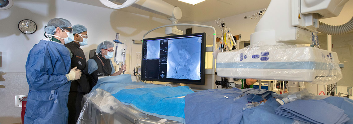
Diseases of the arteries of the lower limbs
Diseases of the arteries of the lower limbs manifest themselves in the form of stenosed segments and blockages of the arteries, the most common cause being atherosclerosis (hardening of the artery).
Symptoms:
They most often manifest as cramps in the calves, hamstrings, and thighs during walking, climbing stairs and running. Such pain subsides after a short rest. With time, the distance of walking begins to shorten, and then the pain also begins to appear during rest. In the next stage, non-healing sores and gangrene will start to appear in the toes. If this condition is not cured the result may be loss (amputation) of a limb or a certain part of it.

Diagnosis and treatment method:
In patients with arterial problems of the lower extremities, it is necessary to first listen to the complaints very well and in detail. Then to check the pulse of these arteries. If necessary, imaging methods are also used. The most commonly applied methods in such a case are colour Doppler, ultrasonography, MRI or CT angiography, and also classical conventional angiography. Although the most commonly used angiography takes the first place here, modern methods of examination such as MRI and CT scanning make it possible to obtain almost perfect results. For this purpose, only a very short, in the range of 15-20 minutes, stay of the patient in the MRI or tomography apparatus is necessary.
There is no need for needle penetration through the groin as in the classical method, and contrasting of the whole body is achieved by intravenous infusion of the required drug through the arm. The electronically obtained images of the required body part are converted into an angiography. Treatment decisions are made using this method. The method of treatment is chosen according to the patient's condition and may involve medication, endovascular therapy, or open surgery.
Blockage of veins
With each beat, the heart sends a certain amount of blood to the tissues and arteries to meet the needs of the body. This clean blood carries oxygen and nutrients to the cells and collects the waste substances formed there. The color of this blood is now dark and it is carried through the veins back to the heart. Varicose veins are in fact dilations along these very veins.
Superficial veins:
They are located immediately under the skin and are visible to the naked eye. We can think of them as the side pathways in our body for carrying contaminated blood to the heart. The most common disease in superficial veins is varicose veins.
Deep veins:
These are veins located adjacent to the arteries, therefore not visible to the naked eye. We can define them as highways, providing the transport of 90% of the dirty blood from the legs to the heart. The most common deep vein diseases are blockages and valvular insufficiency, known as internal varicose veins.
The job of the venous system is to carry blood collected from the tissues to the heart. Unlike the arteries, the veins have very sensitive valves that, despite gravity, ensure the one-way movement of blood to the heart and prevent it from going back the other way. This movement continues to take place even when a person is upright. People forced to stand for prolonged periods on their feet, the function of these valves may be impaired and varicose veins may subsequently appear. Teachers, nurses, surgeons, police officers, dentists and waiters are in the risk group. And vice versa, athletes who also stand on their feet for long periods but use their calf muscles very frequently are less likely to develop varicose veins. Despite it is lower, there is also a risk with prolonged sitting. The rate of varicose veins in occupations such as secretaries, computer operators, etc. is not small.
Interventional radiology in cancer treatment
Interventional radiology as a specialty is a degree higher than classical radiology. In classical radiology, specialists diagnose diseases using methods of visualizing the human body such as radiography, ultrasonography, topography, and MRI. Interventional radiologists, after undergoing special additional training, using the same equipment can now not only diagnose but also treat diseases. Here, no surgical intervention is required; treatment is carried out simply by penetrating the skin with needles or by angiography through blood vessels. Interventional radiology has contributed greatly to the results achieved in recent years in the field of oncology and cancer treatment.
Interventional cancer therapy is a method that has emerged as an alternative to surgery or chemotherapy for certain cancers. Here, treatment is performed as in other types of interventional treatment by the assistance of imaging (e.g. angiography, CT or ultrasonography). The intervention is therefore not a surgery. All interventions are carried out under topical anaesthesia by penetration through a very small opening. Some do not even require a prolonged hospital stay. After several hours of monitoring, the patient can leave the hospital. Others may require a one-day stay and very rarely more. Interventional radiology may also be used for some liver cancers or metastases of other cancers in the liver, for some lung cancers or recurrences in the lungs of other cancers, and for some kidney and bone tumours.
Aneurysm treatment
Aneurysm:
The most common medical formulation of an aneurysm is a pathological dilatation in the form of a bubble in a blood vessel. Aneurysms are most commonly found in the arteries of the brain and the aorta. Aneurysms according to their morphology are saccular and fusiform. Aneurysms occurring at a specific location on the venous wall and having a relatively round shape are called saccular. Spindle-shaped dilatations are called fusiform.
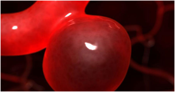
On the other hand, according to their wall structure, aneurysms are divided into "true" and "false or pseudo aneurysms" The wall structure of a true aneurysm includes all the layers normally forming the artery (intima, media, and adventitia). A pseudoaneurysm is formed after a rupture of the wall of a blood vessel and the wall does not contain all the venous layers.
The cerebral arteries and the aorta are the most common sites of location of aneurysms. The incidence of cerebral aneurysms in the general population is 2-3%. Patients diagnosed with polycystic kidney disease and their relatives, people who have a history of subarachnoid hemorrhage due to aneurysm, carry a higher risk of cerebral aneurysm formation.
The structure of the aneurysm wall, compared to the structure of a normal artery, is weaker, which is why it can be ruptured (torn). The rupture can cause life-threatening profuse hemorrhage into the so-called subarachnoid space, limited by the meninges and brain tissue. Such haemorrhages are termed subarachnoid haemorrhages. Unfortunately, the majority of cerebral aneurysms do not give symptoms before a rupture. In a significant part of the patients with aneurysm, the diagnosis is made incidentally during diagnostic imaging such as magnetic resonance imaging (MRI), computed tomography (CT) due to other complaints.
But along with this, large aneurysms may present with complaints and symptoms such as headache, feeling of pain in the fundus of the eye, nausea and vomiting, dropping of the eyelid, double or foggy vision. In general, the annual risk of hemorrhage of a previously unruptured aneurysm is 1-2%. The most common complaint of patients with previous subarachnoid haemorrhage due to aneurysm is sudden onset severe headache. They characterize the phenomenon as "the strongest headache they have had up to that point". In subarachnoid haemorrhage, other common symptoms and complaints, along with headache, are nausea and vomiting, vision blurring or loss of consciousness.
Treatment of brain aneurysms:
In patients who have had a cerebral haemorrhage due to an aneurysm rupture, if the aneurysm is not healed (closed) then in 35% of them within 30 days after the first haemorrhage the aneurysm will rupture once more and become the cause of a second haemorrhage. Therefore, patients with an aneurysm haemorrhage need to be treated as soon as possible. The treatment decision for patients diagnosed with an aneurysm is made considering the age, location and size of the aneurysm and other patient-specific medical criteria. The basic principle for aneurysm treatment is to eliminate the likelihood of rupture (recurrence) of the aneurysm by preventing blood from entering it.
Centre for treatment of varicose veins
What are varicose veins?
Varicose veins are a disease of the human circulatory system, most often occurring in the veins, and especially - the veins of the legs. Varicose veins on the lower extremities most often appear in the area immediately below and above the knee. Due to damage to the valves in this area, a functional disturbance in the system occurs.

Why do varicose veins form?
The veins of the legs perform the function of carrying the dirty blood from the legs upwards back to the heart. At the point above the knee, there are 5-7 valves that prevent the return of the dirty blood directed downwards due to gravity. These valves open and close, thereby preventing dirty blood from mixing with clean blood. But due to some reasons the valves may be damaged and the dirty blood returns down the legs and stays there. This remaining blood causes the veins to dilate and become folded under the skin. The so-called varicose veins form or venous disease occurs.
Many of the health problems at birth are passed on genetically, so if your parents suffered from a venous disease, you will find yourself in a risk group. Especially if your mother had varicose veins the danger in you is increased twice. Of course, the only cause of varicose veins is not heredity.
An important cause of valve damage is intravenous pressure. It is formed due to long immobilization with standing up on your feet. For example, imagine a hairdresser - all day working on his feet, almost without moving. And if for 30-60 minutes he does not move, does not lie down or does not lift his legs to a height of 20-30 cm, then the pressure formed will not go down and will become the cause of damage to the valve. We will move, walk, lie down and even lying down we will raise our legs 20-30 cm up, putting pillows under them so that the pressure formed in the veins will drop.
What are the symptoms of varicose veins, how will we recognize the disease?
During the initial stage, the disease does not have symptoms, but a person will be able to understand when it appears on his own. Especially in later stages, the veins enlarge visibly and can be seen under the skin. This is the most obvious symptom, but the sufferer may also have some difficulties and complaints.
As the veins fold under the skin, especially at night, itching and pain on the legs may occur. The problem of cramp formation on the legs is also common. In case of pain in the legs, placing them 20-30 cm higher on a cushion while sitting and staying like that for a while will ease the pain.
The case is not limited to superficial dilation of veins, after a certain period of time, varicose veins start to burst. This is the so-called bleeding of the varicose vein. The condition is serious enough, so that sometimes stopping the bleeding requires a tamponade on the site. Bleeding can become the cause of non-healing sores on the legs or so-called varicose ulcers. Treatment of varicose ulcers causing necrosis on the legs is the most difficult part in treatment.
What should be done to prevent varicose veins?
Ask your parents if they have varicose disease, and if have they undergone treatment for it. And if their answer is positive, it is necessary to take precautions. The first thing that can be done is to avoid standing still. If your job involves standing on your feet, then every 30-60 minutes provide yourself with movement in the form of a short walk, moving away from the place or if you can lie down for a while. Of course, when the prerequisite is genetic measures cannot always prevent the manifestation.
How are varicose veins treated?
Complete cure of varicose vein is performed by surgical intervention, but there are surgical and non-surgical ways of treatment.
Non-surgical methods: There are 4 methods. Two of them, laser treatment and radiofrequency treatment are subcutaneous and the other two, with foam and sclerotherapy - by infusing medication into the vein using a syringe. The treatment of very thin capillaries, in which needle penetration is impossible, it is carried out supracutaneously.
Surgical treatment: Although today it is considered a rather primitive method, in health care facilities where the necessary equipment and vascular surgeons are not available, unfortunately, it is still used. The operation is performed under complete anaesthesia. A 3 to 7 days hospital stay and 20-45 days of home rest are required.
Interventional radiology in dialysis
On vascular access sites in hemodialysis, the most common pathologies involving the draining veins are anastomosis and stenosis. The most common cause for loss of the vascular access is thrombotic complications.
An irreplaceable method for the evaluation of haemodialysis vascular access in terms of complications are angiographic studies (contrast fistulography and venography). Another method for detecting complications is colour Doppler ultrasonography, which is a non-invasive and relatively inexpensive method, but it depends on the experience and ability of the specialist. colour Doppler ultrasonography has a high sensitivity and is used in the evaluation of vascular access, the arterial system, and in the detection of pseudoaneurysms.
The percutaneous interventional radiological procedures (RTA, stent implantation, catheter repositioning, removal of fibrin sheath of catheters) applied in the treatment of hemodialysis vascular access complications are not only shortening the patient's stay in the hospital, but also providing active and effective treatment methods.
Vascular access (fistula) in dialysis includes native arteriovenous fistulas, arteriovenous grafts and permanent or temporary venous catheters. Vascular access in dialysis patients is considered their lifeline. For a long-term dialysis, a very well-functioning vascular access is of particular importance. The most common reason for hospitalization of patients on hemodialysis is decreased function of this access.
For this reason, it is of great importance for both the patient and the physician to prevent vascular access complications and to diagnose and treat them promptly if they occur. Poor functioning or non-functioning of the vascular access in these patients impedes a normal dialysis program. The occurrence of such a condition necessitates that nephrologists (166) seek another vascular access route in order to continue the hemodialysis process.